Tuesday, December 15, 2015

Can rubisco be used in a technology for carbon capture?

How much rubisco is needed to capture 37 Gigatonnes of CO2 in one year? 37 Gigatonnes is the current amount of human emissions per year.

Assuming that 1 single rubisco enzyme fixes 3 CO2 molecules per second, then in a year 1 mole of rubisco, weighing about 0.49 tonnes, could fix 4163.69 tonnes of CO2. To fix 37 Gigatonnes of CO2 in one year, it will be necessary 4.35 Megatonnes of rubisco. To produce this amount of rubisco in one year, the rate of production needed is 0.138 tonnes of rubisco per second.
I found somewhere that in nature about 1000 tonnes of rubisco are produced per second, so the rate of production would be equivalent to 0.01% of nature’s.

In comparison 300 Megatonnes of plastic are produced each year as of 2013, equivalent to a rate of production of 9.51 tonnes per second.


If you spread the production of 0.138 tonnes of rubisco per second across countries all over the world, then it doesn't really seem like much.

The above example assumed that 1 rubisco could be active for an entire year, but in reality the half-life of rubisco is of several days, which is actually pretty stable. This means the production rate has to be higher than those values to compensate for the inactivation of the enzyme. On the other hand, there are also better rubiscos that can fix more than 3 CO2 molecules per second, so it all balances itself out.

The big question is: can a hybrid system that combines genetic engineer, materials, surfaces, and other industrial technologies be implemented to use the reactions of the Calvin-Benson-Bassham cycle as a
CO2 sequestration mechanism? My colleagues at Imperial would probably say NO… but I’m hopeful that we’re only scratching the surface of the technologies that we’ll be able to develop in the near and far future. This system would have to be fully powered by renewables of course (e.g. solar) and it would have to be better than biomass accumulation.

What do you think?

rubisco
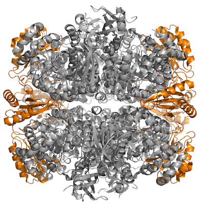
Nature's carbon capture enzyme, also known as rubisco

Tuesday, November 10, 2015

A cyanobacterium with an anoxygenic Type II reaction center from purple bacteria? (Contamination)

With the surge of genome sequences it is crazy what you can find. As I was doing a few BLASTs searching for new photosynthetic reaction center proteins in the database I noticed that the genome of Lyngbya confervoides BDU141951, which was reported early this year (link), contained genes that encode an anoxygenic Type II reaction center from Alphaproteobacteria (Purple Bacteria).

The three genes for the L, M and H subunit.

NameAccessionStartStopStrandGeneIDLocusLocus tagProtein productLengthProtein name
UnNZ_JTHE01000129.114472301+--QQ91_RS05655WP_039723522.1284photosynthetic reaction center subunit L
UnNZ_JTHE01000129.123153298+--QQ91_RS05660WP_039723523.1327photosynthetic reaction center subunit M
UnNZ_JTHE01000254.130083799---QQ91_RS10590WP_039724487.1263photosynthetic reaction center subunit H


1. The best hit for the L subunit was to:

Photosynthetic reaction center subunit L [Oceanibaculum indicum]
ref|WP_008944428.1|

ScoreExpectMethodIdentitiesPositivesGaps
442 bits(1137) 2e-153 Compositional matrix adjust. 222/280(79%) 240/280(85%) 1/280(0%)


2. The best hit for the M subunit was to:

Photosynthetic reaction center subunit M [Ahrensia sp. R2A130]
ref|WP_009758337.1|


ScoreExpectMethodIdentitiesPositivesGaps
457 bits(1175) 5e-158 Compositional matrix adjust. 221/308(72%) 260/308(84%) 2/308(0%)


3. The best hit for the H subunit was to:

Photosynthetic reaction centre, H-chain [Hoeflea phototrophica]
ref|WP_007196606.1|

ScoreExpectMethodIdentitiesPositivesGaps
282 bits(721) 5e-91 Compositional matrix adjust. 139/258(54%) 175/258(67%) 2/258(0%)

What is going on?

When I BLASTed the BchZ subunit of chlorophyllide a reductase from Rhodobacter sphaeroides, which makes bacteriochlorophyllide a from chlorophyllide a... the best hit was to this same strain of cyanobacteria: but it seems it is a fragment only (see image below, never mind the annotation). A close homologous to BchY did not retrieve anything out of the ordinary, the same for BchX. So this cyanobacterium cannot really make bacteriochlorophyll... I wonder if the anoxygenic Type II reaction center in this strain could be active using chlorophyll a along side PSII and PSI.

The fact that the best hits to the reaction center proteins are below 80% means that the alphaproteobacterium that donated this genes represents at least a new genus or something.


UPDATE: It turned out after all to be contamination. It might be that the cultures used were not completely axenic. It's a pity.

Friday, October 30, 2015

Horizontal gene transfer from gammaproteobacteria to cyanobacteria and others

It seems to me that Gammaproteobacteria can transfer a lot of their photosynthesis related genes to other bacteria.

Recent phylogenetic studies (Bryant and Liu, 2013; Sousa et al., 2013) showed that an ancestor of marine Synechococcus and Prochlorococcus strains obtained a set of bchLNB genes from gammaproteobacteria. Such an event of lateral gene transfer from a gammaproteobacterium into an ancestor of the Synechococcus/Prochlorococcus group also included other genes, encoding proteins such as CmpA involved in circadian output (Dvornyk, 2006), carboxisome proteins, rubisco (Marin et al., 2007), threonyl tRNA synthetase, and quite possibly many more (Zhaxybayeva et al., 2006; Zhaxybayeva et al., 2009).

Gemmatimonas photoautotrophica acquired photosynthesis via horizontal gene transfer from a gammaproteobacterium (Zeng et al., 2014) and the same seems true for a Firmicutes of the genus Alkalibacterium. I wonder why...

Microbial mat

References
Bryant, D.A., and Liu, Z.F. (2013). Green bacteria: Insights into green bacterial evolution through genomic analyses. Advances in Botanical Research 66, 99-150.

Dvornyk, V. (2006). Subfamilies of cpmA, a gene involved in circadian output, have different evolutionary histories in cyanobacteria. Microbioliology 152, 75-84.

Marin, B., Nowack, E.C.M., Glockner, G., and Melkonian, M. (2007). The ancestor of the Paulinella chromatophore obtained a carboxysomal operon by horizontal gene transfer from a Nitrococcus-like gamma-proteobacterium. BMC evolutionary biology 7.

Perreault, N.N., Greer, C.W., Andersen, D.T., Tille, S., Lacrampe-Couloume, G., Lollar, B.S., and Whyte, L.G. (2008). Heterotrophic and autotrophic microbial populations in cold perennial springs of the high arctic. Appl. Environ. Microb. 74, 6898-6907.

Sousa, F.L., Shavit-Grievink, L., Allen, J.F., and Martin, W.F. (2013). Chlorophyll biosynthesis gene evolution indicates photosystem gene duplication, not photosystem merger, at the origin of oxygenic photosynthesis. Genome biology and evolution 5, 200-216.

Zeng, Y.H., Feng, F.Y., Medova, H., Dean, J., and Koblizek, M. (2014). Functional Type 2 photosynthetic reaction centers found in the rare bacterial phylum Gemmatimonadetes. P. Natl. Acad. Sci. U. S. A. 111, 7795-7800.

Zhaxybayeva, O., Doolittle, W.F., Papke, R.T., and Gogarten, J.P. (2009). Intertwined evolutionary histories of marine Synechococcus and Prochlorococcus marinus. Genome biology and evolution 1, 325-339.

Zhaxybayeva, O., Gogarten, J.P., Charlebois, R.L., Doolittle, W.F., and Papke, R.T. (2006). Phylogenetic analyses of cyanobacterial genomes: Quantification of horizontal gene transfer events. Genome Research 16, 1099-1108.

Thursday, October 29, 2015

Evolution of oxygenic photosynthesis - New D1 sequences

Early this year I published a study on the evolution of all D1 proteins (Cardona et al., 2015). Interestingly, there are a number of sequences that were very atypical and appeared to be early evolving. Because of their phylogenetic position and sequence characteristics, I suggested that it is possible that these D1 sequences evolved before the water oxidizing complex had reached its standard configuration in PSII.

Back then there were about 40-45 atypical sequences. Since them more have appeared, there are a total of 62 sequences... so here I show a Maximum Likelihood tree for all the atypical D1 forms. See Figure 1. Of particular interests is the fact that they seem to follow an evolutionary pattern consistent with vertical descent and loss, although some likely events of later gene transfer can also be identified.

cyanobacteria evolition oxygenic photosynthesis
Figure 1. Updated tree of atypical D1 sequences.
Of particular interest is the appearance of numerous D1 fragments. Some of them are from incompletely sequenced genes, but some of them seem to be legitimate proteins, probably originating from partial gene duplication followed by divergence (Figure 2). A couple of these fragmented D1 seems to have phylogenetic affinity for the early evolving forms.

oxygenic photosynthesis cyanobacteria
Figure 2. Sequence alignments of different D1. That marked with Fr. is the D1 fragment, found in the genome of some of the earliest evolving cyanobacteria strains, Synechococcus sp. PCC 7336. It seems to have some affinity for the G0 and early branching forms.

Thursday, July 30, 2015

Is a 'tree of life' also a 'tree of death'?

Imagine that you could build an evolutionary tree that included every single organism that has ever existed since the origin of life: including every single bacterium, every single archaeum: every individual organism.

The divisions between groups of organisms would then become blurred. For example, it would be impossible to tell where Homo neanderthalensis ends and where Homo sapiens begins. It would be impossible to detect the exact moment when non-avian dinosaurs turned into birds. This also applies at the unicellular level.

The reason we can distinguish groups in the trees of life that represent evolutionary events, is because we do not see most of the branches: the branches that have gone extinct. It is the absence of these individuals, which did not successfully passed on their genomes to the next generation, that allows for the classification and distinction of different types of organisms.

Therefore, a phylogenetic tree of organisms not only gives insight into the groups of organisms in question, but it also shed lights into the organisms that must have existed but did not make it. You could say that a tree of life is also a tree of death.

It also implies that using a phylogenetic tree we could calculate the amount of diversity that has been lost between the two most closely related branches. Assuming that we know the rate of divergence, and that we could somehow put a number to “amount of diversity”.

extinction evolution
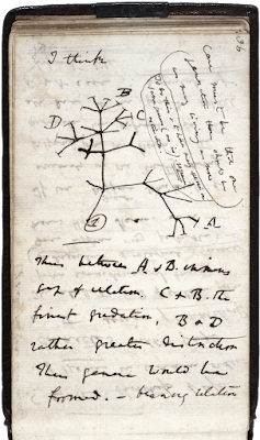
Darwin's Tree of Life, 1837

Monday, July 20, 2015

Phototrophy in ancient Actinobacteria

The evolution of photosynthesis is complex, but not intractable. Views on the origin of photosynthesis can be summarized like this:

Photosynthesis evolved in a well-defined clade of bacteria and then it was scattered through the bacterial tree of life via horizontal gene transfer.

I have come to the conclusion based on the phylogenies of reaction centers, chlorophyll synthesis, and bacteria, that photosynthesis originated close to the origin of bacteria or even before the last common ancestor to bacteria (Cardona, 2014).

A recent paper has shown that two strains of non-phototrophic bacteria of the genus Rubrobacter (phylum: Actinobacteria) contain proteins closely related to those in the chlorophyll-synthesis pathway (Gupta and Khadka, 2015). In addition, the authors show that the origin of these genes is very ancient and provide some evidence to argue that they were not obtained via horizontal gene transfer. They suggest that the Actinobacteria might have been originally phototrophic.

If we reexamine the tree of life and highlight the phototrophic clades (see Figure 1), we see that Cyanobacteria, Chloroflexi, and Actinobacteria likely shared a common ancestor. This relationship is well supported by phylogenomics from the past decade. I believe this ancestor was phototrophic.

photosystem origin evolution
Figure 1. Tree of life highlighting phototrophic clades. The tree is taken from Segata et al., 2013, it's open access and freely available.
In these phylogenetic trees the phylum Firmicutes always branches before the divergence of the Cyanobacteria-Chloroflexi-Actinobacteria supergroup, see Cardona (2014) and references within. This implies that the origin of phototrophy must have predated the divergence of the Firmicutes and the Cyno-Chloro-Actino supergroup, immediately placing the origin of phototrophy very close to the root of bacteria.

Wednesday, April 22, 2015

Contamination of genome projects with DNA from other organisms

I was blasting a protein named PsbO, also known as the 'manganese stabilizing protein' of Photosystem II. This is a protein found in cyanobacteria, algae, and plants, and it is important in photosynthesis. I was doing a phylogenetic tree and noted that one of the proteins originated from the recently sequenced non-photosynthetic bacterium Paenibacillus sp. IHB B 3415. A BLAST showed that the PsbO in this strain is identical to that in Camellia sinensis, the tea plant.

The chances of horizontal gene transfer from the chloroplast of the tea plant to Paenibacillus, I would say, is pretty close to 0%. So I imagine this is some form of contamination.

It is interesting that some of the investigators involved in the genome project are from Hill Area Tea Science Division, CSIR-Institute of Himalayan Bioresource Technology in Palampur, India.

I'm a little bit concerned. What is the chance of contamination to be present in genome projects? In the case of contamination from Eukaryote DNA into that of a bacterium, I guess it is not such a big deal because it can be easily spotted... but if you have contamination from another strain of bacteria, this might look like horizontal gene transfer and it may be not that simple to differentiate using just bioinformatics.

Update (April 20, 2015)
I contacted GenBank to report the issue, they investigated and this is what they told me:

"The submitter concurs with your assessment, so we have removed the contaminated contig JUEI01000195 from the public record." 

manganese stabilizing protein msp
The PsbO protein of Photosystem II